IVF and assessment of embryo development
Before the embryos are transferred, embryologists regularly monitor their development and assign them to specific categories that determine their quality and readiness for transfer into the uterine cavity. The assessment usually takes into account aspects such as:
- rate of development,
- size and shape of the blastomeres (i.e. embryo cells),
- degree of fragmentation.
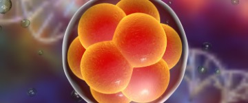
The embryo is assessed during each stage of development, with the exception of Day 4, as this is the compaction stage, during which it is difficult to distinguish individual blastomeres. The following are considered normal:
- Day 2 – the embryo already consists of 4 blastomeres (daughter cells).
- Day 3 – an 8-blastomere embryo is expected; however, each embryo develops individually, and at this stage embryos often have a different number of cells, e.g. 6 or 10, while still remaining within the range of normal development.
The assessment of the morphology of Day 2 and Day 3 embryos takes into account the number of blastomeres and the evaluation of morphological parameters:
- A. – good: <10% fragmentation, blastomere size appropriate for the number of cells, no signs of multinucleation.
- B. – average: 10–25% fragmentation, most embryo cells are of the optimal size for the given stage, no signs of multinucleation.
- C. – poor: >25% fragmentation, considerable variation in cell size, signs of multinucleation present in the blastomeres.
- Day 4 – at the morula stage, a normally developing embryo already consists of more than a dozen cells, which are additionally joined together (the degree of joining, referred to as compaction, is also taken into account during the assessment). At this stage, a three-grade scale is also used.
- Day 5–6 – however, the greatest attention is paid to blastocysts, i.e. embryos on Days 5–6 of development. They are usually assessed using the Gardner method, which involves a six-grade quality classification. A number (from 1 to 6) is used to describe the stage of development they have reached:
- 1 – early blastocyst
- 2 – mature blastocyst
- 3 – expanded blastocyst
- 4 – hatching blastocyst
- 5 – blastocyst undergoing hatching
- 6 – hatched blastocyst
Additionally, they are further classified using letters (A–C), indicating the grade of development and arrangement of the outer cells of the inner cell mass, i.e. the part from which the embryo itself will develop:
- A – many cells tightly packed together
- B – several cells loosely arranged
- C – very few, scattered cells
and the trophectoderm, which will develop into the placenta:
- A – many cells joined together
- B – few cells forming a loose structure
- C – individual cells of relatively large size
Embryo classification in the ivf procedure
Blastocysts classified as AA, AB, BA, and BB are of the highest quality and have the greatest potential for implantation. Embryos classified as CB or BC also have a chance of developing and may be transferred and vitrified. It is important to remember that every embryo is different, and its assessment should always be carried out by embryologists. No diagnoses should be made independently.